Patient guide
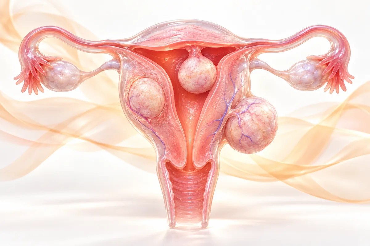
Fibroid types explained: intramural, subserosal and submucosal
How intramural, subserosal and submucosal fibroids differ, the symptoms each tends to cause, how they are diagnosed, and which treatments usually fit which type.
Fibroids at a glance
- 3 typesIntramural, subserosal, submucosal
- MRI firstConfirms type, number and position
- No cutsUFE works through a thin tube
- Day careUFE is usually same-day
- Uterus keptWith embolization


Watch Dr. Nagateja explain this
Short videos on the same topic, in plain language.
The three main types
Fibroids start in the muscle layer of the uterus and grow in different directions.
| Type | Where it grows | Symptoms often seen | Treatment thinking |
|---|---|---|---|
| Intramural | Inside the muscle wall | Heavy periods, bulky uterus, pressure | UFE or myomectomy depending on size and plans [2, 4] |
| Subserosal | On the outer surface | Pressure on bladder or bowel, backache, normal periods | UFE if the base is broad, surgery for narrow stalks |
| Submucosal | Into the uterine cavity | Very heavy bleeding, anaemia, fertility problems | Often hysteroscopic removal, UFE in selected cases [3] |
Symptoms, in plain terms
Common symptoms across all types, with severity depending on size, number and position:
- Heavy or prolonged periods, sometimes with clots
- Tiredness or breathlessness from anaemia
- Pelvic pressure, fullness, or a bulky lower abdomen
- Frequent urination or difficulty emptying the bladder
- Constipation or backache
- Pain during sex, or period pain
How the type is confirmed
A pelvic ultrasound usually finds fibroids. An MRI shows exact size, number and position, including whether a fibroid reaches into the cavity or hangs from a stalk. It also helps tell a fibroid from adenomyosis, which can look similar and is treated differently. MRI is commonly done before UFE.
Which treatments fit which type
These are general patterns. The final plan depends on your age, symptoms, and whether you want a future pregnancy.
Intramural and multiple fibroids respond well to UFE because the blood supply to all of them can be blocked in one session [2, 4]. Subserosal fibroids that sit on a broad base can also be treated this way, while those hanging from a thin stalk usually go to surgery. Submucosal fibroids that push into the cavity are often removed through a hysteroscope, with UFE considered in selected cases [3].
When to book a consultation
See a specialist if your periods are heavy enough to cause tiredness or force you to change protection every hour or two, if you have constant pelvic pressure, or if fibroids were found and you want to understand your choices before agreeing to surgery.
Frequently asked questions
- Which fibroid type is most common?
- Intramural fibroids, inside the muscle wall, are the most common type [1].
- Can fibroids shrink on their own?
- They often shrink after menopause because they depend on hormones. Before menopause they usually stay the same or grow slowly [3].
- Does a large fibroid always need surgery?
- No. Size alone does not decide treatment. Symptoms, position and your plans do. UFE has been used for large fibroids as well, with the suitability assessed on MRI [2, 4].
- What is the difference between fibroids and adenomyosis?
- Fibroids are separate growths in the muscle. Adenomyosis is when the uterine lining tissue grows into the muscle wall, making the whole uterus enlarged and tender. They can coexist and an MRI helps tell them apart.
References
- Society of Interventional Radiology. Uterine fibroids: patient information. Read the source
- RadiologyInfo.org (RSNA and ACR). Uterine fibroid embolization (UFE). Read the source
- NHS. Fibroids: treatment. Read the source
- Johns Hopkins Medicine. Uterine artery embolization. Read the source
Want to discuss your options?
Speak with the Clinic Seven team.