Vascular Malformation Treatment in Hyderabad Complex vessels.
Thoughtful care.
Image-guided options tailored to the location and type of malformation.
Care shaped around your condition.
A detailed assessment guides treatment for abnormal blood vessels.
Image-guided embolization and sclerotherapy treat abnormal clusters of vessels, reducing pain, swelling, bleeding risk and visible deformity.
About Vascular Malformations
Vascular malformations are abnormal collections of veins, arteries, lymphatic channels or mixed vessels that a person is born with, though many are noticed only in the teenage or adult years when they cause pain, swelling, bleeding, skin changes or cosmetic concern. At Clinic Seven in Hyderabad, an Interventional Radiologist uses MRI and Doppler ultrasound to classify the malformation using the ISSVA framework, then treats it with image-guided sclerotherapy or embolization through a small needle or catheter access.
Low-flow venous malformations and lymphatic malformations respond well to sclerotherapy, while high-flow arteriovenous malformations (AVMs) need staged embolization with coils, glue or particles. Treatment aims to shrink the abnormal vessels, relieve pain and swelling, reduce bleeding risk, and improve cosmetic appearance while sparing nearby nerves, skin and normal vessels.
- MRI and Doppler-based ISSVA classification for accurate treatment planning.
- Sclerotherapy for low-flow venous and lymphatic malformations.
- Staged embolization for high-flow arteriovenous malformations.
- Reduces pain, swelling, bleeding risk and cosmetic deformity.




Personalized scenarios are presented during consultation. Individual results may vary.
Symptoms of Vascular Malformations (venous, lymphatic, AVM)
Vascular malformations are present from birth but often become noticeable later. Symptoms depend on the type and location:
- A soft swelling that enlarges when you bend or strain (venous)
- A bluish or reddish patch or lump under the skin
- Pain, heaviness or throbbing over the area
- A warm lump with a pulse or thrill (arteriovenous)
- Recurrent swelling or fluid leaks (lymphatic)
- Bleeding or skin ulceration over the lesion
- Limb overgrowth or asymmetry
- Cosmetic concern on the face or limbs
If these symptoms sound familiar, book an evaluation with Dr. Nagateja Bonala at Clinic Seven, Hyderabad.
- •Tiny, symptomless lesions safely kept under observation
- •Infantile haemangiomas that regress on their own
- •Lesions where surgery is clearly better positioned
- •Active local infection until treated
How the Vascular Malformations procedure works
Every step is planned and performed under image guidance by an Interventional Radiologist at Clinic Seven, Hyderabad.
- 01
Classify
MRI, Doppler and clinical review identify the malformation type.
- 02
Plan
The safest route and staged treatment strategy are selected.
- 03
Treat
Sclerotherapy or embolization closes the abnormal vessels.
- 04
Review
Follow-up tracks shrinkage, symptom relief and whether another stage is needed.
Sclerotherapy & embolization vs open surgical excision
| Factor | Vascular Malformations | Open surgical excision |
|---|---|---|
| Access | Needle or catheter, image-guided | Open dissection |
| Anaesthesia | Local or general by extent | General |
| Normal tissue | Preserved | Risk to nearby nerves and skin |
| Bleeding risk | Low and controlled | High for AVMs |
| Staged treatment | Yes, safely | Difficult, often incomplete |
| Recurrence | Managed with repeat sessions | Common if incomplete |
The right choice depends on your scans, symptoms and goals. Your Interventional Radiologist will talk you through both options at your consultation.
Recovery timeline
Most patients treated at Clinic Seven follow a predictable recovery path with a follow-up review scheduled at the first visit.
- Swelling may last a few days.
- Compression if advised.
- Staged sessions may be needed.
Risks and safety
Every procedure carries some risk. These are the ones your Interventional Radiologist will review before Vascular Malformations.
- Temporary swelling, bruising or skin irritation.
- Rare nerve, skin or vessel injury depending on location.
- Incomplete response may require staged sessions.
What affects the cost
The cost of sclerotherapy or embolization for a vascular malformation in Hyderabad depends on the type, the size and the number of sessions needed. Symptomatic malformations are covered by many insurance plans; our team helps confirm eligibility.
For a personalised estimate and insurance guidance, contact Clinic Seven with your recent reports.
- Type of malformation (venous, lymphatic or AVM)
- Number of staged sessions
- Sclerosant, coils or glue used
- MRI and Doppler workup
- Hospital and room category
- Insurance and cashless approval
Get a rough idea of the cost
Choose what matters to you and we will show a guide. Nobody is committed to anything, and our team will never rush you.
Is your insurance accepted?
Cashless with major insurers, TPAs and office group policies. CGHS and Singareni on credit.
Popular searches
Cover is confirmed by our team after checking your policy and treatment plan.
View all insurersDr. Nagateja Bonala
Dr. Nagateja Bonala is the Interventional Radiologist at Clinic Seven, Hyderabad, with 13+ years of image-guided practice. Every Vascular Malformations case is planned personally with a review of your scans and reports.
Plan your next step
Bring your recent scans, blood tests and any prior specialist notes. Message us on the contact page for directions, timings and insurance guidance.
Questions about Vascular Malformations
A vascular malformation is an abnormal cluster of blood or lymphatic vessels present from birth. It can involve veins (venous malformation), lymph channels (lymphatic malformation), arteries and veins together (arteriovenous malformation) or a mix, and can appear anywhere in the body.
Infantile hemangiomas grow in the first year of life and shrink on their own over years. Vascular malformations do not spontaneously shrink and often enlarge slowly with growth, hormonal changes, trauma or infection.
Clinical examination, Doppler ultrasound and, for anything more than superficial, MRI are the mainstay. Classification follows the ISSVA framework, which guides treatment strategy.
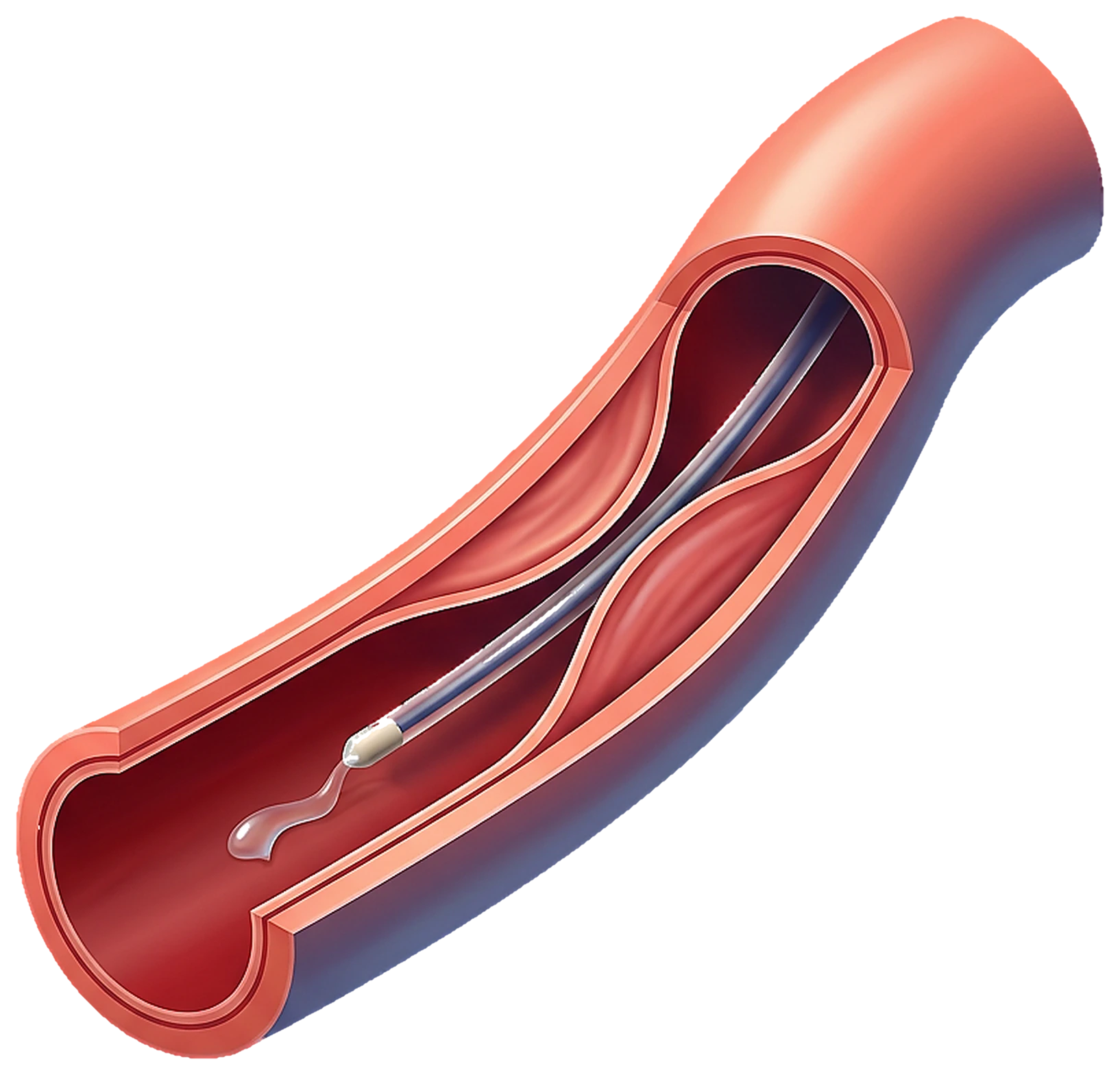
A medicine called a sclerosant is injected directly into the abnormal vein or lymph channel through a small needle under ultrasound or X-ray guidance. It inflames the lining of the vessel so it seals shut and shrinks over weeks.
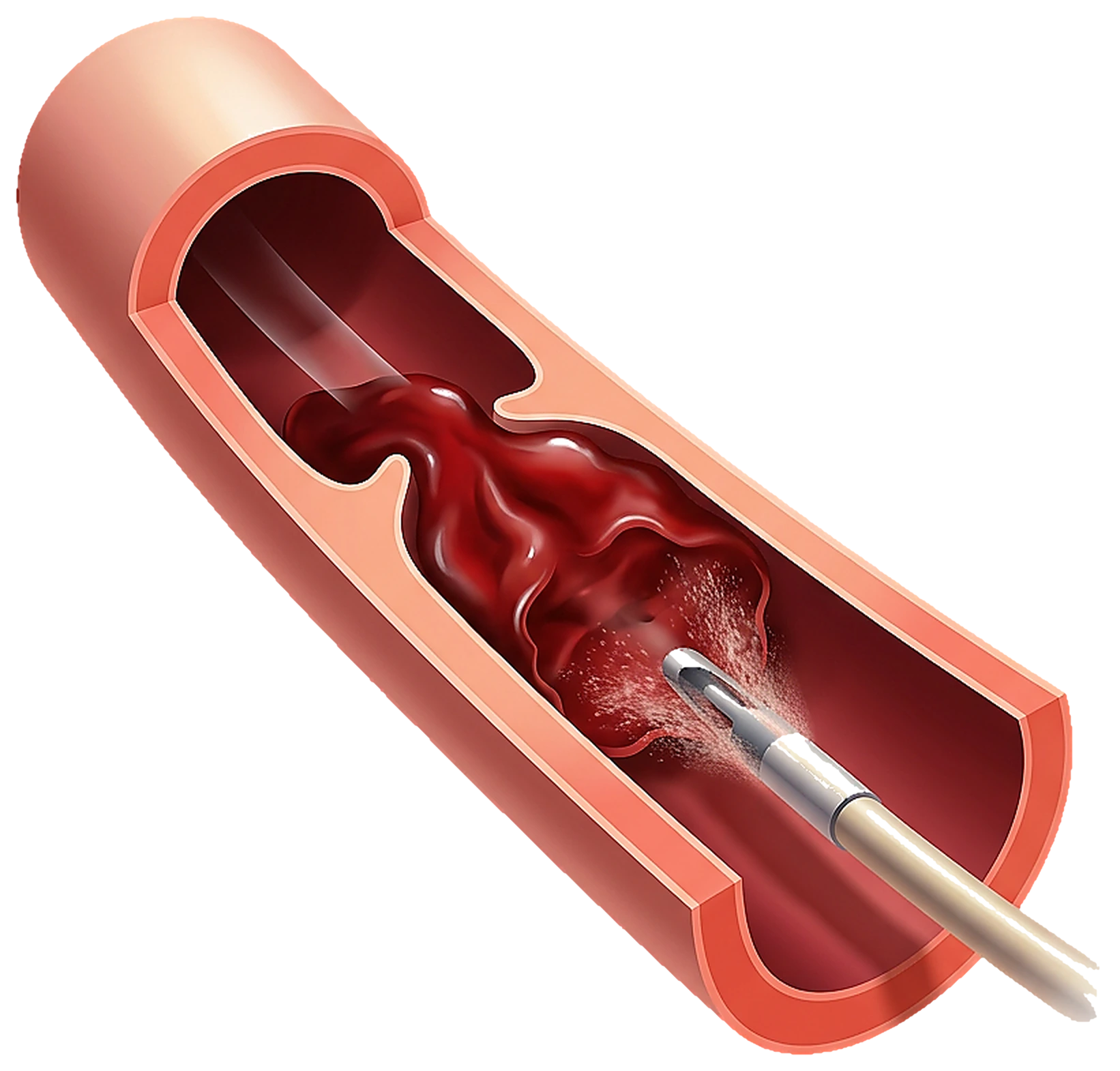
In an AVM, a catheter is guided into the nidus of abnormal vessels and closed off with coils, glue or particles. Staged sessions are often needed to safely close all feeding vessels.
Some small malformations respond to a single session. Extensive or complex lesions are usually planned in stages to protect surrounding skin, nerves and muscle, and to allow safe response between sessions.
Many are only cosmetic. Others can bleed, ulcerate, hurt, cause limb overgrowth, or, in high-flow AVMs, place strain on the heart. Early evaluation by an experienced team is important.
Clinic Seven, Hyderabad offers image-guided sclerotherapy and embolization for vascular malformations by Dr. Nagateja Bonala, coordinated with plastic surgery, dermatology and paediatric teams when needed.
Related treatments at Clinic Seven
Minimally invasive, image-guided options by Dr. Nagateja Bonala in Hyderabad.