Patient guide
Breast lump biopsy: ultrasound-guided core biopsy explained
How a breast lump is biopsied under ultrasound, why a core biopsy is usually preferred to FNAC, what the studies show, marker clips, aftercare and how long results take.
Breast biopsy at a glance
- Ultrasound guidedNeedle seen throughout
- Local anaestheticYou stay awake
- A small punctureNo stitches
- Day-careHome the same day
- A clear diagnosisTissue for the pathologist
- Quick recoveryBack to routine soon



When a breast lump needs a biopsy
Most breast lumps turn out to be benign, such as fibroadenomas or cysts. Even so, a lump that is new, hard, growing, or that looks suspicious on imaging should be checked with a needle biopsy. The standard approach is the combination of examination, imaging (ultrasound, mammography or MRI) and a biopsy, so that the diagnosis does not rest on any one test.
If the biopsy shows a benign lump such as a fibroadenoma, you may be able to simply monitor it, and there are non-surgical treatment options if it is large or troublesome.
Why ultrasound-guided core biopsy is usually chosen
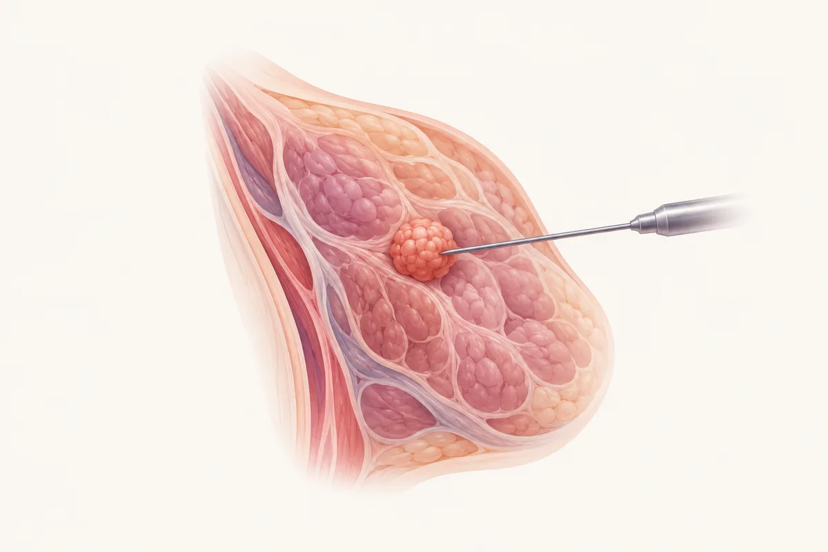
A core needle takes thin cores of tissue, so the pathologist sees the architecture of the lump and can run special tests, including the receptor tests (ER, PR and HER2) that guide breast cancer treatment. FNAC collects only loose cells, which cannot give the same information. Ultrasound lets the doctor watch the needle enter the lump, so the sample comes from the right part and the armpit glands can be sampled in the same sitting.
Winkler et al., J Breast Imaging 2023, 140 patients, same node sampled both ways [2].
How the biopsy is done
Most breast biopsies take about 20 to 30 minutes in all.
- You lie on your back or slightly turned, and the skin is cleaned.
- Local anaesthetic numbs the skin and the tissue around the lump.
- Using ultrasound, the doctor guides a core needle into the lump and takes several cores. You hear a click, and feel pressure.
- A tiny marker clip may be left at the site, which shows on later scans.
- Pressure is held, and a dressing is placed. You go home the same day.
Aftercare and risks
Bruising and tenderness for a few days are common. A supportive bra and simple pain relief help. Avoid heavy lifting or vigorous exercise for the period you are advised. Bleeding or a larger bruise (haematoma) and infection are uncommon, and the team will tell you what to watch for. Tell the team about blood thinners beforehand and do not stop them without instructions [1].
Results and next steps
A first report usually takes a few days, and receptor tests take longer if cancer is found. Your doctor explains the result and the plan. A benign result that does not match the imaging may lead to a repeat or a larger sample, and a cancer result leads to a coordinated plan with your breast surgeon and oncologist.
Cost and insurance
Health insurance can cover these procedures, subject to your policy terms. The exact cost is calculated after consultation, a complete evaluation of the problem, and a decision on the procedure that suits you. Cashless treatment can be arranged with major Indian health insurers and TPAs, and credit treatment is available for CGHS and Singareni beneficiaries. Eligibility is confirmed after a policy check.
Frequently asked questions
- Is a breast biopsy painful?
- The area is numbed first. You may feel pressure and a click. Soreness for a few days is usual.
- Does a breast biopsy cause cancer to spread?
- No. Needle biopsy is the standard way to diagnose breast lumps, and seeding along the needle track is extremely rare.
- FNAC or core biopsy for a breast lump?
- Core biopsy usually gives more information, including receptor tests if cancer is found, and was more sensitive for axillary nodes in studies [2]. FNAC may still be used in some situations. The doctor advises after seeing your imaging.
- What is a marker clip?
- A tiny metal clip that marks the biopsy site on later scans. It is safe, and it helps the surgeon find the area if surgery is needed.
- Can a fibroadenoma be treated without surgery?
- In selected women, yes. A biopsy confirms the diagnosis first, and there are image-guided options for fibroadenomas. See the fibroadenoma treatment page.
References
- Patel IJ, Rahim S, Davidson JC, et al. Society of Interventional Radiology Consensus Guidelines for the Periprocedural Management of Thrombotic and Bleeding Risk in Patients Undergoing Percutaneous Image-Guided Interventions, Part II: Recommendations. J Vasc Interv Radiol. 2019;30(8):1168-1184. Read the source
- Winkler N, Buxton J, Freer P, et al. Comparison of diagnostic sensitivity and procedure-related pain of concurrent ultrasound-guided fine-needle aspiration and core-needle biopsy of axillary lymph nodes in patients with suspected or known breast cancer. J Breast Imaging. 2023;5(4):436-444. Read the source
Want to discuss your options?
Speak with the Clinic Seven team.