Patient guide
Ultrasound-guided biopsy: how it works, safety and what to expect
What an ultrasound-guided biopsy is, which lumps and organs it is used for, how it is done, how safe it is, preparation and when CT guidance is used instead.
Ultrasound-guided biopsy at a glance
- Needle seen liveSteered to the right spot
- Local anaestheticNo general anaesthesia
- A small punctureA dressing, no stitches
- Walk in, walk outUsually day-care
- About 15-30 minutesFor most biopsies
- No radiationSound waves only


What an ultrasound-guided biopsy is
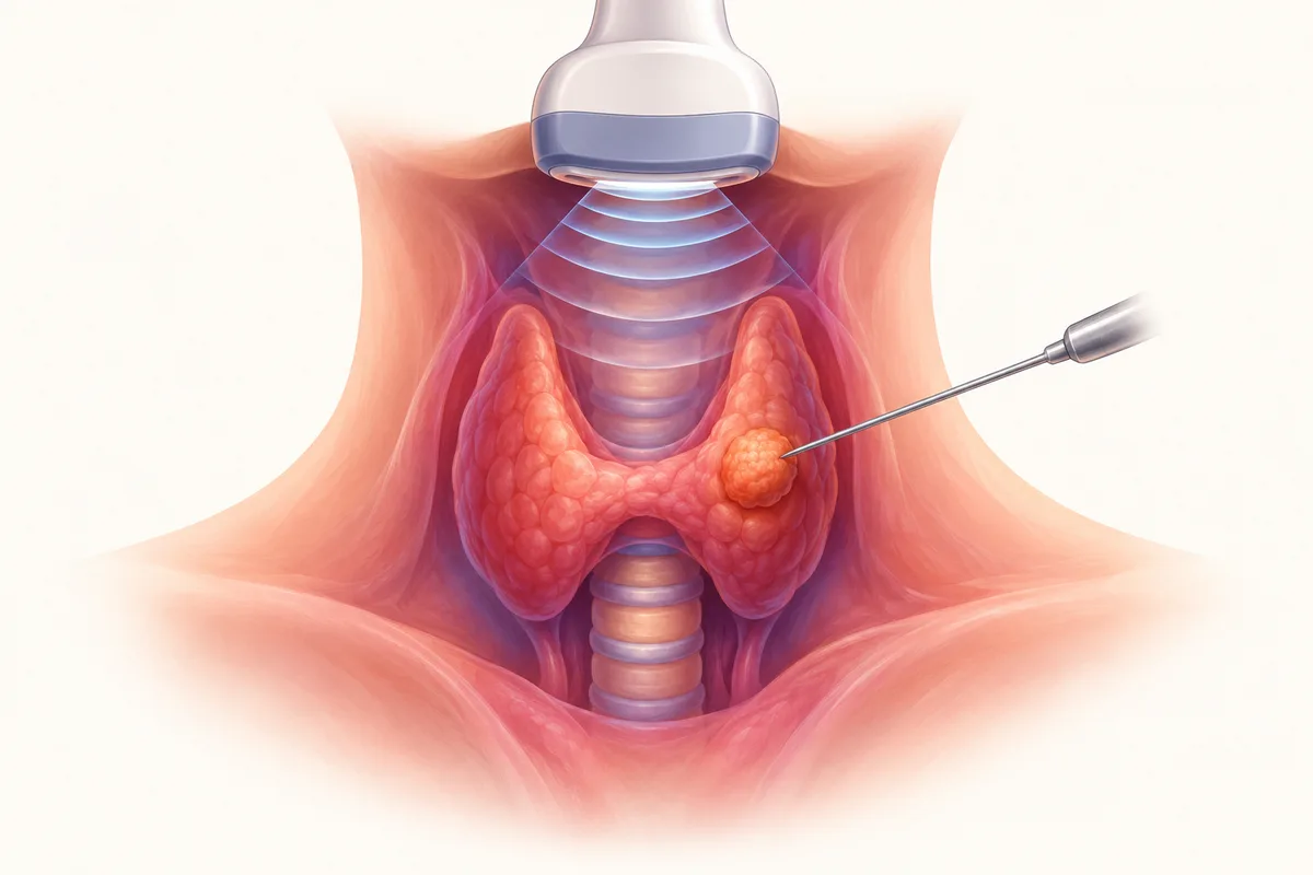
A biopsy takes a small sample from a lump or organ so that a pathologist can examine it. When the needle is guided by ultrasound, the doctor watches the needle tip on the screen as it enters the target, so the sample comes from the right part of the abnormality and nearby structures such as vessels are avoided.
The sample can be taken with a very fine needle (FNAC, which collects cells) or a slightly wider needle that removes a thin core of tissue (a core or Trucut biopsy). Both can be done under ultrasound, and the choice depends on the target and the question being asked.
What it is used for
Ultrasound is the usual guide for anything it can see clearly through the skin.
- Thyroid nodules, including the decision about whether a nodule needs treatment.
- Enlarged glands in the neck, armpit or groin.
- Breast lumps and axillary glands.
- Liver, kidney and other abdominal masses that are reachable.
- Soft-tissue lumps under the skin.
- Fluid collections, which can be sampled or drained in the same sitting.
How it is done
Most ultrasound-guided biopsies take about 15 to 30 minutes in total, including preparation.
- You lie in the position that gives the best view. The skin is cleaned and a sterile drape is placed.
- The skin and tissue along the needle path are numbed with local anaesthetic.
- The doctor guides the needle into the target under live ultrasound and takes one or more samples. A core needle makes a short clicking sound.
- Pressure is held on the spot, and a small dressing is placed. There are no stitches.
- You are watched for a short while, and usually go home the same day.
How safe it is
Ultrasound-guided biopsy is generally very safe, and the risk depends on the organ. In a systematic review of 39 studies and 14,818 patients, the pooled complication rate after ultrasound-guided core biopsy of thyroid nodules was 1.11%, and major complications were about 0.06% [2]. The usual issues are a bruise, a little local bleeding or discomfort, and they settle on their own.
Bleeding risk is the main thing planned around. Superficial biopsies are generally lower risk, and deeper organ biopsies such as liver or kidney carry a higher bleeding risk and need blood tests and a plan for blood thinners [1].
Ha et al., Eur Radiol 2018, 39 studies, 14,818 patients [2].
When CT is used instead
Ultrasound waves do not pass through air or bone. Targets in the lung, behind the bowel, deep in the abdomen or in bone are often better reached with CT guidance. If the doctor judges that ultrasound cannot show a target safely, you will be told and the biopsy will be planned with CT instead.
Preparing and what happens next
Bring your scans and reports. Tell the team about blood thinners and other medicines, and do not stop any on your own. You may be asked to fast for liver or kidney biopsies. After the procedure, avoid heavy lifting or strenuous activity for the period you are advised, keep the dressing dry, and watch for swelling, fever or bleeding.
The sample goes to the laboratory, and your doctor explains the report and the next step when it is ready.
Cost and insurance
Health insurance can cover these procedures, subject to your policy terms. The exact cost is calculated after consultation, a complete evaluation of the problem, and a decision on the procedure that suits you. Cashless treatment can be arranged with major Indian health insurers and TPAs, and credit treatment is available for CGHS and Singareni beneficiaries. Eligibility is confirmed after a policy check.
Frequently asked questions
- Is ultrasound-guided biopsy painful?
- The area is numbed with local anaesthetic. You may feel a pinch and pressure. Most people find it uncomfortable at worst.
- What is the difference between USG-guided FNAC and a USG-guided core biopsy?
- FNAC uses a very fine needle to collect cells, while a core biopsy uses a wider needle to remove a thin piece of tissue, which gives the pathologist more to examine. The right choice depends on the target [3].
- Does an ultrasound-guided biopsy spread cancer?
- Needle-track seeding is described but very uncommon, and the value of knowing the diagnosis far outweighs it. Your doctor will weigh this for your particular situation.
- Can I eat before an ultrasound-guided biopsy?
- For thyroid, breast and neck biopsies you can usually eat normally. For liver or kidney biopsies you may be asked to fast for a few hours. Follow the instructions given when it is booked.
- How soon can I resume normal activity?
- Most people resume light activity the same day. Avoid heavy lifting or strenuous exercise for as long as you are advised.
References
- Patel IJ, Rahim S, Davidson JC, et al. Society of Interventional Radiology Consensus Guidelines for the Periprocedural Management of Thrombotic and Bleeding Risk in Patients Undergoing Percutaneous Image-Guided Interventions, Part II: Recommendations. J Vasc Interv Radiol. 2019;30(8):1168-1184. Read the source
- Ha EJ, Suh CH, Baek JH. Complications following ultrasound-guided core needle biopsy of thyroid nodules: a systematic review and meta-analysis. Eur Radiol. 2018;28(9):3848-3860. Read the source
- Zhao Q, Meng Q, Wang Z. Diagnostic performance and operator workload of core needle biopsy versus fine-needle aspiration for superficial lymph nodes: a prospective, paired comparative study. Acad Radiol. 2026;33(8):3425-3435. Read the source
Want to discuss your options?
Speak with the Clinic Seven team.