Patient guide
CT-guided biopsy: lung, abdomen and deep lesions explained
What a CT-guided biopsy is, when it is used (lung nodules, deep masses, bone), how it is done, the risk of pneumothorax and bleeding, preparation and recovery.
CT-guided biopsy at a glance
- Pinpoint accuracyThe needle is checked on CT slices
- Local anaestheticYou stay awake
- A small punctureA dressing, no stitches
- Short observationMany go home the same day
- Reaches deep targetsLung, pancreas, bone and more
- About 30-60 minutesFor most CT biopsies



What a CT-guided biopsy is
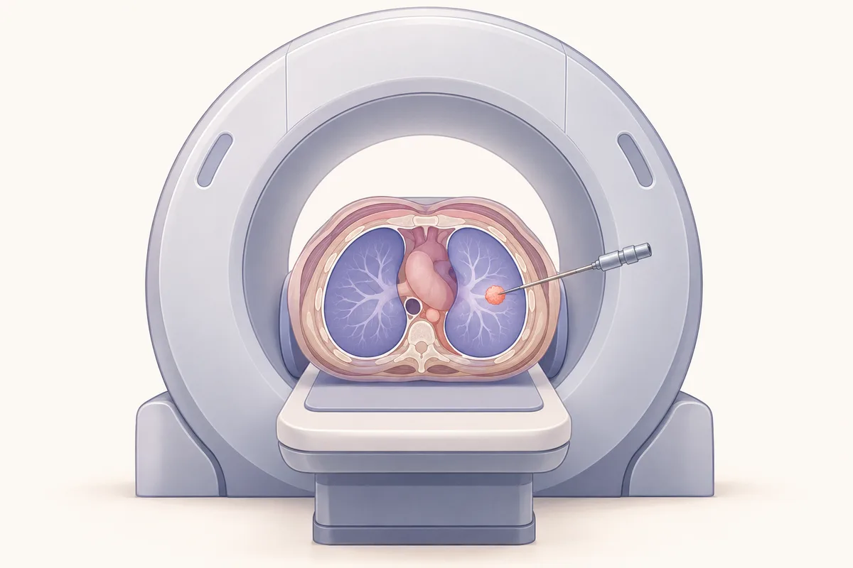
CT shows the body in thin cross-sections, so it can guide a needle to a target that ultrasound cannot reach or see well. The doctor plans the safest path on the scan, numbs the skin, advances the needle in steps with repeat scans, and takes a sample once the tip is confirmed in the target.
The sample can be cells (FNAC) or a thin core of tissue (a core or Trucut biopsy), and the pathologist then identifies what the lesion is.
When CT guidance is used
CT is chosen when ultrasound cannot show the target safely.
- Lung nodules, masses and shadows, including those near the chest wall or the middle of the chest (mediastinum).
- Deep abdominal and retroperitoneal masses, the pancreas and adrenal glands.
- Enlarged deep glands in the chest or abdomen.
- Bone and soft-tissue lesions near the spine or pelvis.
- Lesions that need a very precise path, close to major vessels or other structures.
How it is done
Plan around 30 to 60 minutes for most CT-guided biopsies, including preparation and the scans.
- A scan is done first, and the doctor plans the safest route and marks the skin.
- You lie in the position that gives the best path, often on your back, side or front, and you are asked to keep still and breathe as instructed.
- The skin and tissue are numbed with local anaesthetic.
- The needle is advanced in stages with checking scans, and the sample is taken once the tip is confirmed in the target.
- A dressing is placed and you are observed. For lung biopsies, a chest X-ray is done after a few hours to check for a pneumothorax.
Risks: pneumothorax and bleeding
For lung biopsies, the main risk is pneumothorax, where a small amount of air leaks between the lung and the chest wall. A small one often settles by itself, and a larger one may need a thin tube to remove the air. A pooled analysis of 32 studies of core biopsy (8,133 procedures) and 17 studies of FNA (4,620 procedures) found overall complication rates of 38.8% for core biopsy and 24.0% for FNA, mostly minor, with major complication rates of 5.7% and 4.4% [2]. In that analysis, FNA complications were more likely with smaller lesions, larger needles and more lung traversed.
Bleeding is also planned for: deep biopsies fall in the higher bleeding risk group, so blood tests are checked and blood thinners are managed with your doctor beforehand [1].
Heerink et al., Eur Radiol 2017, 12,753 procedures [2].
Who may not be suitable, and radiation
A CT-guided biopsy needs you to lie still and follow breathing instructions. Severe lung disease, an uncontrolled bleeding tendency or a target in a very risky position may make the risk higher, and your doctor will discuss alternatives. Radiation from the scans is kept as low as reasonably possible, and in almost all cases the value of a firm diagnosis outweighs it. Tell the team if you may be pregnant.
Preparing and recovery
You may be asked to fast for a few hours. Bring your scans and blood tests, and do not stop blood thinners without instructions. After a lung biopsy you are asked to rest, avoid strenuous activity and flying for the period advised, and report breathlessness, worsening chest pain, coughing blood or fever straight away. Results of tissue samples take from a few days to a week or more when special stains are needed.
Cost and insurance
Health insurance can cover these procedures, subject to your policy terms. The exact cost is calculated after consultation, a complete evaluation of the problem, and a decision on the procedure that suits you. Cashless treatment can be arranged with major Indian health insurers and TPAs, and credit treatment is available for CGHS and Singareni beneficiaries. Eligibility is confirmed after a policy check.
Frequently asked questions
- Is CT-guided biopsy safe?
- It is widely used and the major complication rate is low, at around 4 to 6% in pooled lung biopsy data, but minor problems such as a small pneumothorax are common [2]. Your doctor explains your own risk, which depends on the target and your lungs.
- Will I be put to sleep?
- No. Local anaesthetic is used and you stay awake so you can follow breathing instructions. Light sedation is possible in some cases.
- What is the difference between ultrasound-guided and CT-guided biopsy?
- Ultrasound is used when the target can be seen clearly through the skin, with no radiation. CT is used for deep or hidden targets, such as the lung or pancreas, where precise placement matters.
- Can I go home on the same day?
- Often yes. Lung biopsies need a few hours of observation and a chest X-ray first, and the doctor decides if a longer stay is needed.
- How long do CT-guided biopsy results take?
- Usually a few days, longer when special stains are needed. Your doctor will explain the report and the next step.
References
- Patel IJ, Rahim S, Davidson JC, et al. Society of Interventional Radiology Consensus Guidelines for the Periprocedural Management of Thrombotic and Bleeding Risk in Patients Undergoing Percutaneous Image-Guided Interventions, Part II: Recommendations. J Vasc Interv Radiol. 2019;30(8):1168-1184. Read the source
- Heerink WJ, de Bock GH, de Jonge GJ, et al. Complication rates of CT-guided transthoracic lung biopsy: meta-analysis. Eur Radiol. 2017;27(1):138-148. Read the source
Want to discuss your options?
Speak with the Clinic Seven team.